Lymph nodes are small oval-shaped balls of lymphatic tissue, distributed widely throughout the body and linked by a vast network of lymphatic vessels. Lymph nodes are repositories of B cells, T cells, and other immune system cells, such as dendritic cells and macrophages. They act as filters for foreign particles in the body and are one of the sites where adaptive immune responses are triggered.
Structure of Lymph Nodes
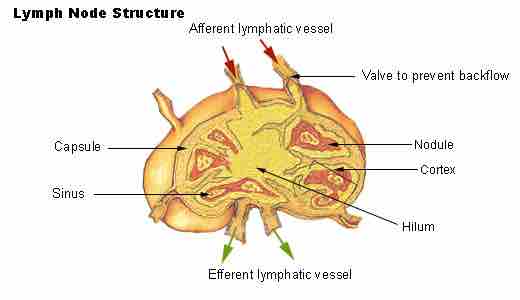
Lymph nodes are found throughout the body, and are typically 1 to 2 centimeters long. Humans have approximately 500-600 lymph nodes, with clusters found in the underarms, groin, neck, chest, and abdomen. Each lymph node is surrounded by a fibrous capsule that encircles the internal cortex and medulla. The cortex is mainly composed of clusters of B cells in the outer layers and T cells in the inner layers, and may also contain antigen-presenting dendritic cells. The medulla contains plasma cells, macrophages, and B cells as well as sinuses, which are vessel-like spaces that the lymph flows into. Inside each sinus cavity is a nodule, a smaller, denser bundle of lymphoid tissue that usually contains a germinal center, the site of B cell proliferation during antigen presentation. The sinuses are partially divided by capsule tissue, which causes lymph fluid to flow around the nodules in each sinus cavity on their way through the node.

Lymph node structure
This diagram of a lymph node shows the outer capsule, cortex, medulla, hilum, sinus, valve to prevent backflow, nodule, and afferent and efferent vessels.

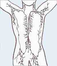
The lymphatic system
This diagram shows the network of lymph nodes and connecting lymphatic vessels in the human body.
Lymph fluid flows into and out of the lymph nodes via the lymphatic vessels, a network of valved vessels that are similar in structure to cardiovascular veins. Each lymph node has an afferent lymph vessel that directs lymph into the node, and an efferent lymph vessel called the hilum that directs lymph out of the node at the concave side of the node. The hilum also contains the blood supply of the lymph node.
Function of Lymph Nodes
Lymph nodes are the primary site for antigen presentation and activation in adaptive immune response in B and T lymphocytes. These lymphocytes are continuously recirculated through the lymph nodes and the bloodstream. Molecules called antigens are found on bacteria cell walls, the cell walls of virus-infected cells, or even chemical substances and toxins secreted from bacteria. These antigens may be taken by cells into the lymph nodes. There, antigen-presenting cells called dendritic cells present the antigen molecule to naive B and T lymphocytes. These undergo cell cycle proliferation into lymphocytes that are able to specifically detect and eliminate pathogens associated with that antigen, through various methods such as cytotoxic action (T cells) and antibody production (B cells).
The lymph nodes also filter the lymph fluid. Macrophages in the sinus spaces phagocytize (engulf) foreign particles such as pathogen, so that lymph fluid that returns to the bloodstream is cleaned of problematic abnormalities. The lymph node is also arranged in such a way that the chance of B and T lymphocytes encountering dendritic cells is quite high, to facilitate antigen presentation.
Lymphadenopathy
Lymphadenopathy describes the clinical condition of swollen lymph nodes. This is usually caused by increased lymph flow into the nodes. This fluid may carry a higher amount of debris, so inflammation occurs as more neutrophils and later macrophages enter the node to remove debris from the lymph.
Lymphadenopathy is a symptom in conditions from trivial, such as a common cold or a minndor infection, to life-threatening, such as cancer or severe infection. Cancers that are severe and widespread from frequent metastases tend to have lymphadenopathy, so cancer staging criteria includes lymph node involvement. Additionally, cancers like lymphomas that have tumors made out of aberrant lymphocytes nearly always show lymphadenopathy, often an early warning sign for this type of cancer.