Chapter 18
Cardiovascular System: Blood Vessels
By Boundless
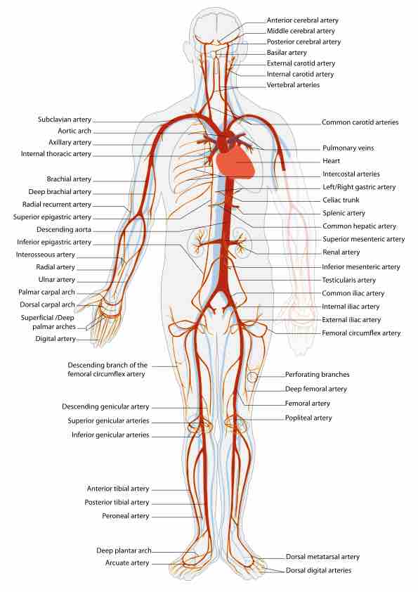
Arteries are high-pressure blood vessels that carry oxygenated blood away from the heart to all other tissues and organs.
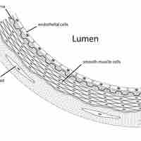
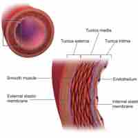
An elastic or conducting artery has a large number of collagen and elastin filaments in the tunica media.
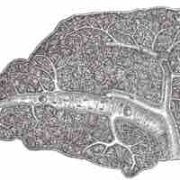
Distributing arteries are medium-sized arteries that draw blood from an elastic artery and branch into resistance vessels.
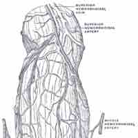
A circulatory anastomosis is a connection or looped interaction between two blood vessels.
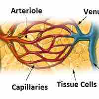
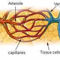
An arteriole is a small diameter blood vessel in the microcirculation system that branches out from an artery and leads to capillaries.
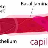
Capillaries, the smallest blood vessels in the body, are part of the microcirculation.
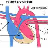
The circulatory system is the continuous system of tubes that pumps blood to tissues and organs throughout the body.
Humans have a closed cardiovascular system, meaning that blood never leaves the network of arteries, veins, and capillaries.
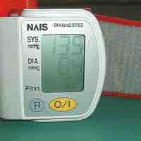
Blood pressure is a vital sign reflecting the pressure exerted on blood vessels when blood is forced out of the heart during contraction.
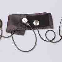
The measurement of blood pressure without further specification usually refers to systemic arterial pressure measured at the upper arm.
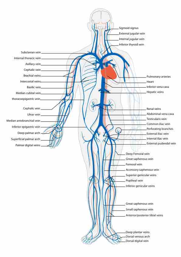
Venous pressure is the vascular pressure in a vein or the atria of the heart, and is much lower than arterial pressure.
The cardiovascular system plays a role in body maintenance by transporting hormones and nutrients and removing waste products.
Neural regulation of blood pressure is achieved through the role of cardiovascular centers and baroreceptor stimulation.
Blood pressure is controlled chemically through dilation or constriction of the blood vessels by vasodilators and vasocontrictors.
Consistent and long-term control of blood pressure is determined by the renin-angiotensin system.
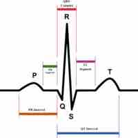
Checking circulation involves measurement of blood pressure and pulse through a variety of invasive and noninvasive methods.
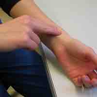
Pulse is a measurement of heart rate by touching and counting beats at several body locations, typically at the wrist radial artery.
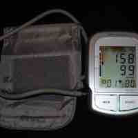
Measurement of blood pressure includes systolic pressure during cardiac contraction and diastolic pressure during cardiac relaxation.
Chronically elevated blood pressure is called hypertension, while chronically low blood pressure is called hypotension.
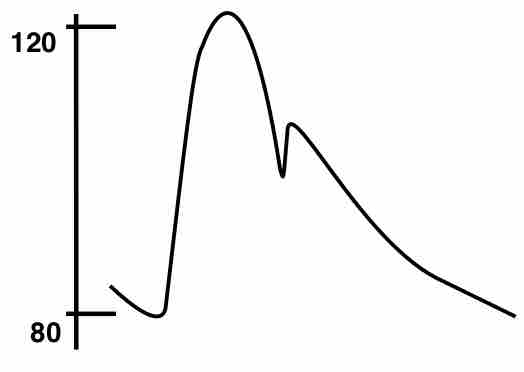
Blood flow is a pulse wave that moves out from the aorta and through the arterial branches, then is reflected back to the heart.
Blood flow is regulated locally in the arterioles and capillaries using smooth muscle contraction, hormones, oxygen, and changes in pH.
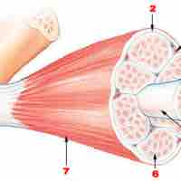
Blood flow to an active muscle changes depending on exercise intensity and contraction frequency and rate.
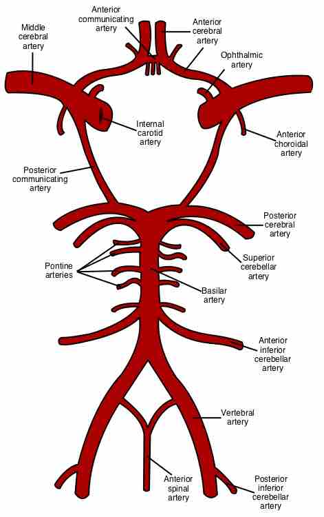
Cerebral circulation is the movement of blood through the network of blood vessels supplying the brain, providing oxygen and nutrients.
Blood flow to the skin provides nutrition to skin and regulates body heat through the constriction and dilation of blood vessels.
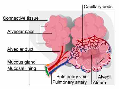
Pulmonary circulation in the lungs is responsible for removing carbon dioxide from and replacing oxygen in deoxygenated blood.
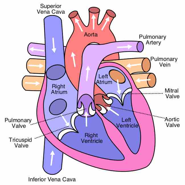
The heart pumps oxygenated blood to the body and deoxygenated blood to the lungs.
The hepatic portal system is responsible for directing blood from parts of the gastrointestinal tract to the liver.
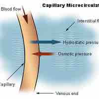
Hydrostatic and osmotic pressure are opposing factors that drive capillary dynamics.
Transcytosis is a process by which molecules are transported into the capillaries.
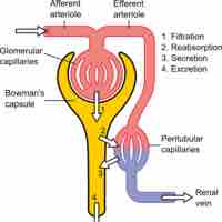
Capillary fluid movement occurs as a result of diffusion (colloid osmotic pressure), transcytosis, and filtration.
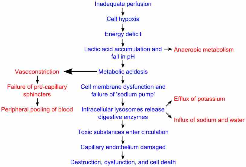
Circulatory shock is a life-threatening medical condition that occurs due to inadequate substrate for aerobic cellular respiration.

An organism responds with numerous reactions during each of the four stages of shock in an attempt to maintain cellular homeostasis.
The clinical manifestation of shock varies depending on the type of shock and the individual, but there are some general symptoms.

The aorta is the largest artery in the body and is divided into 3 parts: the ascending aorta, arch of the aorta, and descending aorta.
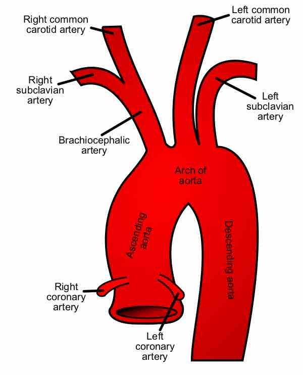
The ascending aorta is the first portion of the aorta; it includes the aortic sinuses, the bulb of the aorta, and the sinotubular junction.

The arch of the aorta follows the ascending aorta and begins at the level of the second sternocostal articulation of the right side.
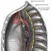
The thoracic aorta is the section of the aorta that travels through the thoracic cavity to carry blood to the head, neck, thorax and arms.
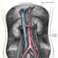
The abdominal aorta is the largest artery in the abdominal cavity and supplies blood to most of the abdominal organs.
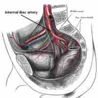
The abdominal aorta divides into the major arteries of the leg: the femoral, popliteal, tibial, dorsal foot, plantar, and fibular arteries.

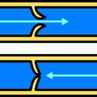
Veins are blood vessels that carry blood towards the heart, have thin, inelastic walls, and contain numerous valves.
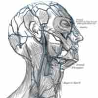
In the head and neck, blood circulates from the upper systemic loop, which originates at the aortic arch.
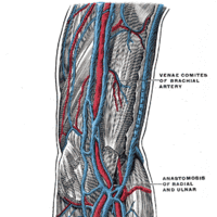
The veins of the upper extremity are divided into superficial and deep veins, indicating their relative depths from the skin.
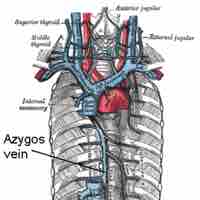
The veins of the thorax drain deoxygenated blood from the thorax region for return to the heart.
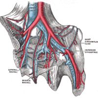
The major veins of the abdomen and pelvis return deoxygenated blood from the abdomen and pelvis to the heart.
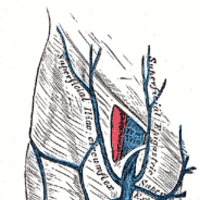
The deep veins of the lower extremity have valves for unidirectional flow and accompany the arteries and their branches.
