A congenital heart defect is a defect in the structure of the heart and great vessels which is present at birth. Many types of heart defects exist, most of which either obstruct blood flow in the heart or vessels near it, or cause blood to flow through the heart in an abnormal pattern. Other defects, such as long QT syndrome, affect the heart's rhythm. Heart defects are among the most common birth defects and are the leading cause of birth defect-related deaths. Approximately nine people in 1,000 are born with a congenital heart defect. Many defects do not require treatment, but some complex congenital heart defects require medication or surgery.
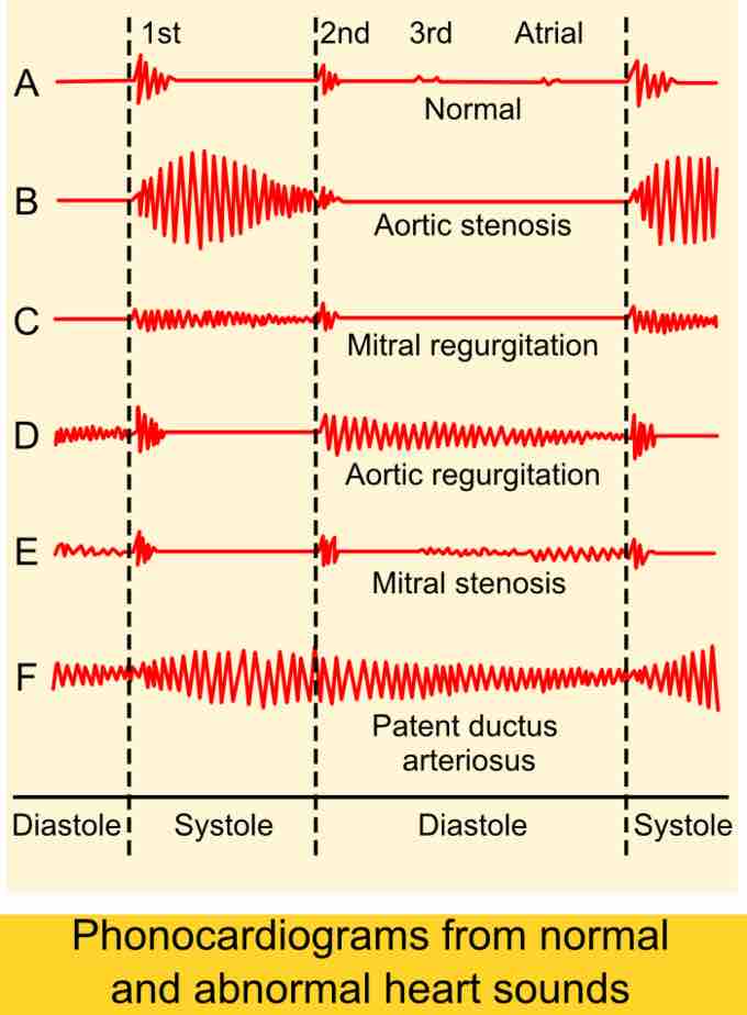
Signs and symptoms are related to the type and severity of the heart defect. Some children have no signs while others may exhibit shortness of breath, cyanosis, syncope, heart murmur, under-developing of limbs and muscles, poor feeding or growth, or respiratory infections. Congenital heart defects cause abnormal heart structure resulting in production of certain sounds called heart murmur. These can sometimes be detected by auscultation ; however, not all heart murmurs are caused by congenital heart defects.

Auscultogram of heart sounds, including murmurs
Changes in heart sounds can indicate specific congenital defects in heart valves and chambers.
There is a complex sequence of events that result in a well formed heart at birth and disruption of any portion may result in a defect. The orderly timing of cell growth, cell migration, and programmed cell death (apoptosis) has been studied extensively and the genes that control the process are being elucidated. In the fetus, the lungs are unexpanded and cannot accommodate the full circulatory volume. Cells in part of the septum primum die creating a hole while muscle cells, the septum secundum, grow along the right atrial side the septum primum, except for one region, leaving a gap, the foramen ovale, through which blood can pass from the right artium to the left atrium, circumventing the pulmonary circuit. A small vessel, the ductus arteriosus allows blood from the pulmonary artery to pass to the aorta. Defects in any of these structures are associated with an increased incidence of other symptoms, together referred to as VACTERL association: V - Vertebral anomalies, A - Anal atresia, C - Cardiovascular anomalies, T - Tracheoesophageal fistula, E - Esophageal atresia, R - Renal (Kidney) and/or radial anomalies, L - Limb defects.
Genetics
Most of the known causes of congenital heart disease are sporadic genetic changes, either focal mutations or deletion or addition of segments of DNA. Small chromosomal abnormalities also frequently lead to congenital heart disease. The genes regulating the complex developmental sequence have only been partly elucidated. Some genes are associated with specific defects. Mutations of a heart muscle protein, α-myosin heavy chain (MYH6) are associated with atrial and septal defects. Several proteins that interact with MYH6 are also associated with cardiac defects. Another factor, the homeobox (developmental) gene, NKX2-5 also interacts with MYH6. Mutations of all these proteins are associated with both atrial and ventricular septal defects.
Fetal Environment
Known antenatal environmental factors that may lead to congenital heart defects include maternal infections (Rubella), drugs (alcohol, hydantoin, lithium, and thalidomide) and maternal illness (diabetes mellitus, phenylketonuria, and systemic lupus erythematosus). As noted in several studies following similar body mass index (BMI) ranges, prepregnant and gestating women, who were obese (BMI ≥ 30), carried a statistically significant risk of birthing children with congenital heart defects (CHD) compared to normal-weight women (BMI= 19-24.9). Although there are minor conflicting reports, there was significant support for the risk of fetal CHD development in overweight mothers (BMI= 25-29.9).
Hypoplasia
Hypoplasia can affect the heart, typically resulting in the underdevelopment of the right ventricle or the left ventricle. This results in only one side of the heart capable of pumping blood to the body and lungs effectively. Hypoplasia of the heart is rare, but is the most serious form of CHD. It is called hypoplastic left heart syndrome when it affects the left side of the heart and hypoplastic right heart syndrome when it affects the right side of the heart. In both conditions, emergency heart surgery will permit blood to circulate to the body (or lungs, depending on which side of the heart is defective). Hypoplasia of the heart is generally a cyanotic heart defect.
Treatment
Sometimes CHD improves without treatment. Other defects are so small that they do not require any treatment. Most of the time, CHD is serious and requires surgery and/or medications. Medications include diuretics, which aid the body in eliminating water, salts, and digoxin for strengthening the contraction of the heart. This slows the heartbeat and removes some fluid from tissues. Interventional cardiology now offers patients minimally invasive alternatives to surgery. Most patients require life-long specialized cardiac care, first with a pediatric cardiologist and later with and adult congenital cardiologist. There are more than 1.8 million adults living with congenital heart defects.

Congenital heart defects
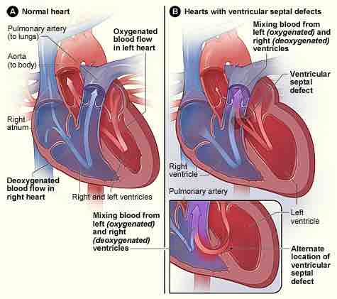
Figure A shows the structure and blood flow in the interior of a normal heart. Figure B shows two common locations for a ventricular septal defect. The defect allows oxygen-rich blood from the left ventricle to mix with oxygen-poor blood in the right ventricle.