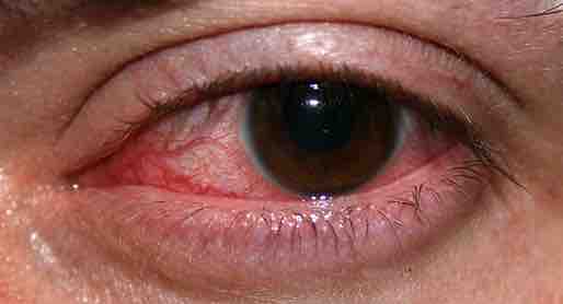
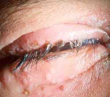
Microbial corneal infection is the most serious and "most common vision threatening" complication of wearing contact lenses, which is believed to be strongly associated with contact lens cases. Such infections "are being increasingly recognized as an important cause of morbidity and blindness" and "may even be life-threatening. " While the cornea is believed to be the most common site for fungal eye infections, other parts of the eye such as the orbit, sclera, and eyelids may also be involved.
Fungal Infections of the Eye
Factors that contribute to fungal contamination of contact lenses include, but not limited to, hygiene negligence such as: improper sterilization and disinfection of contact lenses, use of contaminated lenses, contaminated contact lens case, contaminated contact lens solution, wearing of contact lenses during eye infections and introduction of micro-organisms from the environment.
Diagnosis is determined"by recognition of typical clinical features and through direct microscopic detection of fungi in scrapes, biopsy specimens, and other samples. "Ultimately, cultures that are made from the samples isolated from patients is what "confirms diagnosis. " Other tests that may also be used if needed include "histopathological, immunohistochemical, or DNA-based tests. Pathogenesis of the fungal contaminants includes a wide range of factors such as invasiveness, toxigenicity, and host factors. Once diagnosis is accessed, specific anti-fungal therapy can be administered. One of the most popular and common treatments used"for life-threatening and severe ophthalmic mycoses is amphotericin B which is a specific anti-fungal drug. For the treatment for filamentous fungal keratitis, "topical natamycin is usually the first choice. For the treatment of yeast keratitis, topical amphotericin B is usually the first choice. Current advances in further treatments include evaluations of triazoles such as itraconazole and fluconazole" as therapeutic options in ophthalmic mycoses.

Keratitis
An eye with non-ulcerative sterile keratitis.
Viral Infections of the Eye
Herpes Simplex Virus
Herpetic simplex keratitis is a form of keratitis caused by recurrent herpes simplex virus in cornea. Herpes simplex virus (HSV) infection is very common in humans. HSV is a double-stranded DNA virus that has icosahedral capsid. HSV-1 infections are found more commonly in the oral area and HSV-2 in the genital area. Primary infection most commonly manifests as blepharoconjunctivitis i.e. infection of lids and conjunctiva that heals without scarring. Lid vesicles and conjunctivitis are seen in primary infection. Corneal involvement is rarely seen in primary infection. Recurrent herpes of the eye in turn is caused by reactivation of the virus in a latently infected sensory ganglion, transport of the virus down the nerve axon to sensory nerve endings, and subsequent infection of ocular surface. The following classification of herpes simplex keratitis is important for understanding this disease:

Herpes simplex blepharitis
Primary eye infection with herpes simplex virus most commonly manifests as blepharoconjunctivitis i.e. infection of lids and conjunctiva that heals without scarring. Lid vesicles and conjunctivitis are seen in primary infection.
Dendritic Ulcer (Epithelial Keratitis)
This classic herpetic lesion consists of a linear branching corneal ulcer (dendritic ulcer). During eye exam the defect is examined after staining with fluorescein dye. The underlying corneal has minimal inflammation. Patients with epithelial keratitis complain of foreign-body sensation, light sensitivity, redness, and blurred vision. Focal or diffuse reduction in corneal sensation develops following recurrent epithelial keratitis. In immune deficient patients or with the use of corticosteroids the ulcer may become large and in these cases it is called geographic ulcer.
Disciform Keratitis (Stromal Kieratitis)
Stromal keratitis manifests as a disc-shaped area of corneal edema. Longstanding corneal edema leads to permanent scarring. It is the major cause of decreased vision associated with HSV. Localized endotheliis (localized inflammation of corneal endothelial layer) is the cause of disciform keratitis.
Diagnostic testing is seldom needed because of its classic clinical features and is not useful in stromal keratitis as there is usually no live virus. Laboratory tests are indicated in complicated cases when the clinical diagnosis is uncertain and in all cases of suspected neonatal herpes infection. Corneal smears or impression cytology specimens can be analyzed by culture, antigen detection, or fluorescent antibody testing. Demonstration of HSV is possible with viral culture. Serologic tests in turn may show a rising antibody titer during primary infection but are of no diagnostic assistance during recurrent episodes.
Treatment of herpes of the eye is different based on its presentation. Epithelial keratitis is caused by live virus. Stromal disease is an immune response. Metaherpetic ulcer results from inability of the corneal epithelium to heal. Epithelial keratitis is treated with topical antivirals, which are very effective with low incidence of resistance. Acyclovir ophthalmic ointment and Trifluridine eye drops have similar effectiveness but are more effective than Idoxuridine and Vidarabine eye drops. Topical antiviral medications are not absorbed by the cornea through an intact epithelium, but orally administered acyclovir penetrates an intact cornea and anterior chamber.
Cytomegalovirus Retinitis
Cytomegalovirus retinitis, also known as CMV retinitis, is an inflammation of the retina of the eye that can lead to blindness. Caused by human cytomegalovirus, it occurs predominantly in people whose immune system has been compromised.
Parasitic Infections of the Eye
Acanthamoeba
Acanthamoeba is a microscopic, free-living ameba (single-celled living organism) commonly found in the environment that can cause rare, but severe, eye illness. Acanthamoeba causes three main types of illness involving the eye (Acanthamoeba keratitis), the brain and spinal cord (Granulomatous Encephalitis), and infections that can spread throughout the entire body (disseminated infection).
Toxoplasma gondii
A single-celled parasite called Toxoplasma gondii causes a disease known as toxoplasmosis. While the parasite is found throughout the world, more than 60 million people in the United States may be infected with the Toxoplasma parasite. Of those who are infected, very few have symptoms because a healthy person's immune system usually keeps the parasite from causing illness.
Signs and symptoms of ocular toxoplasmosis can include reduced vision, blurred vision, pain (often with bright light), redness of the eye, and sometimes tearing. Ophthalmologists sometimes prescribe medicine to treat active disease. Whether or not medication is recommended depends on the size of the eye lesion, the location, and the characteristics of the lesion (acute active, versus chronic not progressing). An ophthalmologist will provide the best care for ocular toxoplasmosis.