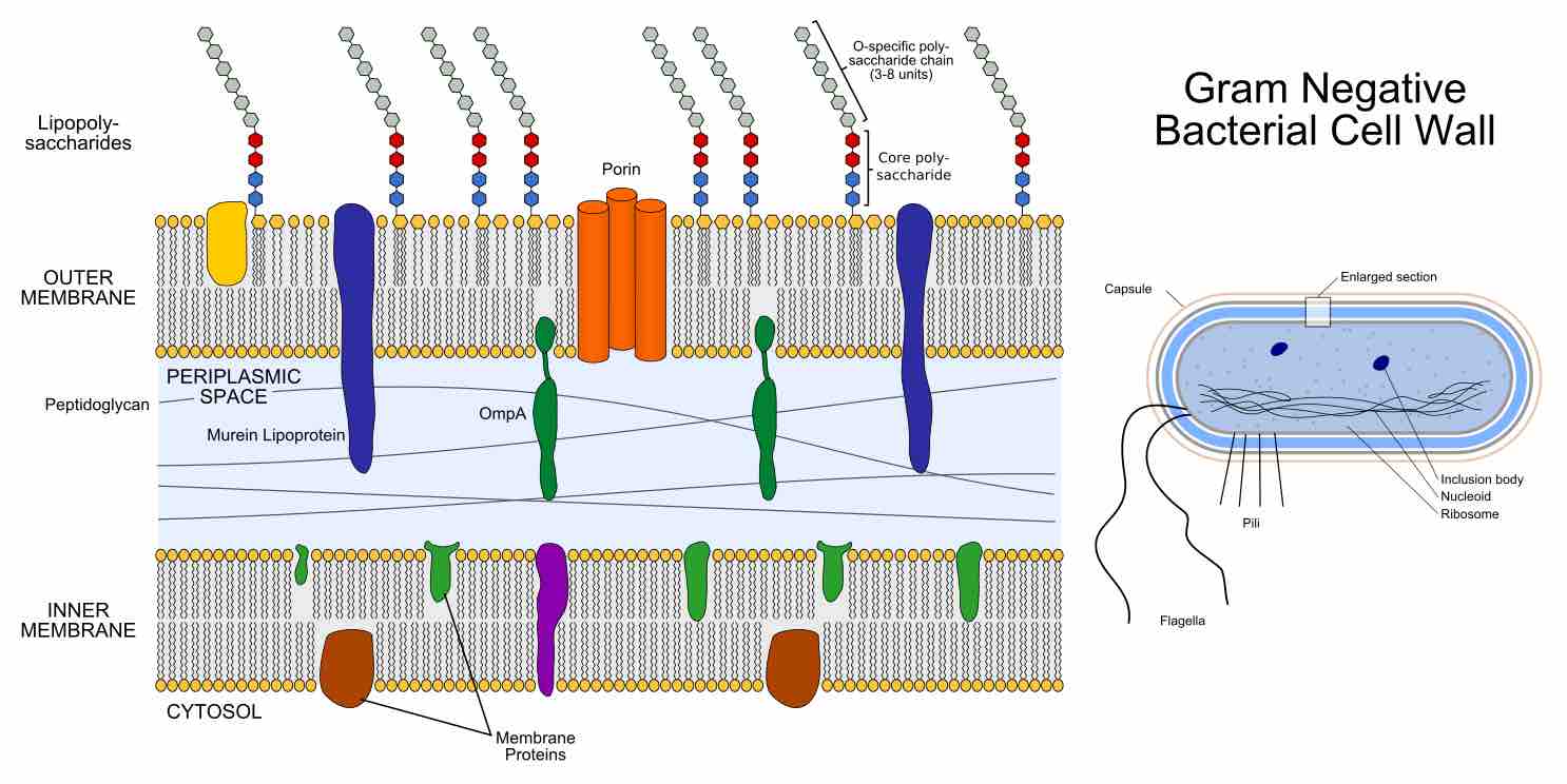
In the Gram-negative Bacteria the cell wall is composed of a single layer of peptidoglycan surrounded by a membranous structure called the outer membrane. The gram-negative bacteria do not retain crystal violet but are able to retain a counterstain, commonly safranin, which is added after the crystal violet. The safranin is responsible for the red or pink color seen with a gram-negative bacteria. The Gram-negative's cell wall is thinner (10 nanometers thick) and less compact than that of Gram-positive bacteria, but remains strong, tough, and elastic to give them shape and protect them against extreme environmental conditions . The outer membrane of Gram-negative bacteria invariably contains a unique component, lipopolysaccharide (LPS) in addition to proteins and phospholipids. The LPS molecule is toxic and is classified as an endotoxin that elicits a strong immune response when the bacteria infect animals.

Structure of Gram-negative cell wall
Gram-negative outer membrane composed of lipopolysaccharides.
In Gram-negative bacteria the outer membrane is usually thought of as part of the outer leaflet of the membrane structure and is relatively permeable. It contains structures that help bacteria adhere to animal cells and cause disease. The peptidoglycan layer is non-covalently anchored to lipoprotein molecules called Braun's lipoproteins through their hydrophobic head. Sandwiched between the outer membrane and the plasma membrane, a concentrated gel-like matrix (the periplasm) is found in the periplasmic space. It is in fact an integral compartment of the gram-negative cell wall and contains binding proteins for amino acids, sugars, vitamins, iron, and enzymes essential for bacterial nutrition. The periplasm space can act as reservoir for virulence factors and a dynamic flux of macromolecules representing the cell's metabolic status and its response to environmental factors. Together, the plasma membrane and the cell wall (outer membrane, peptidoglycan layer, and periplasm) constitute the gram-negative envelope.