Exhaustion is the third and final stage in the general adaptation syndrome model. At this point, all of the body's resources are eventually depleted and the body is unable to maintain normal function. The initial autonomic nervous system symptoms may reappear (sweating, raised heart rate, etc.).
If stage three is extended, long-term damage may result, as the body's immune system becomes exhausted, and bodily functions become impaired and result in de-compensation. The result can manifest itself in obvious illnesses such as ulcers, depression, diabetes, trouble with the digestive system, or even cardiovascular problems, along with other mental illnesses.
Post-Traumatic Stress Disorder
Post-traumatic stress disorder (PTSD) is a severe anxiety disorder that can develop after exposure to any event that results in psychological trauma . Diagnostic symptoms for PTSD include re-experiencing the original trauma(s) through flashbacks or nightmares, avoidance of stimuli associated with the trauma, and increased arousal, such as difficulty falling or staying asleep, anger, and hypervigilance.
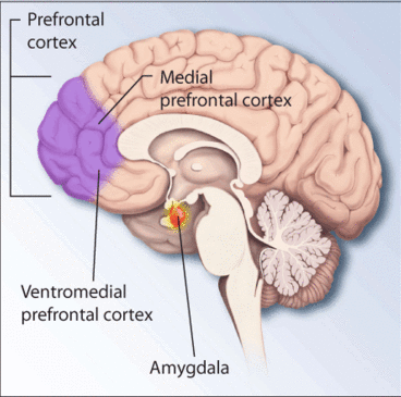
Sensory input, memory formation, and stress response mechanisms are affected in patients with post-traumatic stress disorder. The regions of the brain involved in memory processing that are implicated in PTSD include the hippocampus, amygdala, and frontal cortex, while the heightened stress response is likely to involve the thalamus, hypothalamus, and locus coeruleus.
There is consistent evidence from MRI volumetric studies that hippocampal volume is reduced in post-traumatic stress disorder. This atrophy of the hippocampus is thought to represent decreased neuronal density.
However, other studies suggest that hippocampal changes are explained by whole brain atrophy, and generalized white matter atrophy is exhibited by people with PTSD.

Regions of the brain associated with stress and PTSD
Post-traumatic stress disorder (PTSD) is a severe anxiety disorder that can develop after exposure to psychological trauma.
Memory
Cortisol works with epinephrine (adrenaline) to create memories of short-term emotional events; this is the proposed mechanism for the storage of flash-bulb memories, and may originate as a means to remember what to avoid in the future.
However, long-term exposure to cortisol damages cells in the hippocampus, which results in impaired learning. Furthermore, it has been shown that cortisol inhibits memory retrieval for already stored information.
Depression
Many areas of the brain appear to be involved in depression, including the frontal and temporal lobes and parts of the limbic system, including the cingulate gyrus. However, it is not clear if the changes in these areas cause depression or if the disturbance occurs as a result of the etiology of psychiatric disorders.
The Hypothalamic-Pituitary-Adrenal Axis in Depression
In depression, the hypothalamic-pituitary-adrenal (HPA) axis is up-regulated by a down-regulation of its negative feedback controls. Corticotropin-releasing factor is over-secreted from the hypothalamus and induces the release of adrenocorticotropin hormone (ACTH) from the pituitary.
ACTH interacts with receptors on adrenocortical cells and cortisol is released from the adrenal glands. Adrenal hypertrophy can also occur due to this repeated stimulation. The release of cortisol into the circulatory system has a number of effects, including elevation of blood glucose.
The negative feedback of cortisol to the hypothalamus, pituitary, and immune systems is impaired. This leads to a continual activation of the HPA axis and excess cortisol release. The cortisol receptors then become desensitized, which causes an increase in activity of the pro-inflammatory immune mediators and disturbances in neurotransmitter transmission.
Serotonin Pathways in Depression
Serotonin transmission from both the caudal raphe nuclei and rostral raphe nuclei is reduced in patients with depression compared with non-depressed controls. Increasing the levels of serotonin in these pathways by reducing serotonin re-uptake, hence increasing serotonin function, is one of the therapeutic approaches to treating depression.
The Noradrenaline Pathways in Depression
In depression, the transmission of noradrenaline is reduced from both of the principal noradrenergic centres. An increase in noradrenaline in the frontal/prefrontal cortex modulates the action of selective noradrenaline re-uptake inhibition and improves mood. Increasing noradrenaline transmission to other areas of the frontal cortex modulates attention.
Heart Disease
Excessive cortisol release also has a negative impact on heart health. High levels of cortisol correlate with an increased risk of heart disease. This is due to the increases in blood sugar and blood pressure levels that cortisol imparts along with it's pro-inflammatory effects.