Definition of Carboxysomes
Carboxysomes are intracellular structures found in many autotrophic bacteria, including Cyanobacteria, Knallgasbacteria, Nitroso- and Nitrobacteria. They are proteinaceous structures resembling phage heads in their morphology; they contain the enzymes of carbon dioxide fixation in these organisms. It is thought that the high local concentration of the enzymes, along with the fast conversion of bicarbonate to carbon dioxide by carbonic anhydrase, allows faster and more efficient carbon dioxide fixation than is possible inside the cytoplasm. Similar structures are known to harbor the B12-containing coenzyme glycerol dehydratase, the key enzyme of glycerol fermentation to 1,3-propanediol, in some Enterobacteriaceae, such as Salmonella.
Carboxysomes are bacterial microcompartments that contain enzymes involved in carbon fixation. Carboxysomes are made of polyhedral protein shells about 80 to 140 nanometres in diameter. These compartments are thought to concentrate carbon dioxide to overcome the inefficiency of RuBisCo (ribulose bisphosphate carboxylase/oxygenase) - the predominant enzyme in carbon fixation and the rate limiting enzyme in the Calvin cycle. These organelles are found in all cyanobacteria and many chemotrophic bacteria that fix carbon dioxide.
Carboxysomes are an example of a wider group of protein micro-compartments that have dissimilar functions but similar structures, based on homology of the two shell protein families. Using electron microscopy the first carboxysomes were seen in 1956, in the cyanobacterium Phormidium uncinatum. In the early 1960s, similar polyhedral objects were observed in other cyanobacteria. These structures were named polyhedral bodies in 1961; over the next few years they were also discovered in some chemotrophic bacteria that fixed carbon dioxide. Among these are Halothiobacillus, Acidithiobacillus, Nitrobacter and Nitrococcus.
Carboxysomes were first purified from Thiobacillus neapolitanus in 1973, and were shown to contain RuBisCo held within a rigid outer covering .

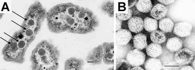
Electron Micrograph of a carboxysome
(A) A thin-section electron micrograph of H. neapolitanus cells with carboxysomes inside. In one of the cells shown, arrows highlight the visible carboxysomes. (B) A negatively stained image of intact carboxysomes isolated from H. neapolitanus. The features visualized arise from the distribution of stain around proteins forming the shell as well as around the RuBisCO molecules that fill the carboxysome interior. Scale bars indicate 100 nm.