Concept
Version 6
Created by Boundless
Carboxysomes

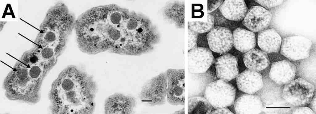
Electron Micrograph of a carboxysome
(A) A thin-section electron micrograph of H. neapolitanus cells with carboxysomes inside. In one of the cells shown, arrows highlight the visible carboxysomes. (B) A negatively stained image of intact carboxysomes isolated from H. neapolitanus. The features visualized arise from the distribution of stain around proteins forming the shell as well as around the RuBisCO molecules that fill the carboxysome interior. Scale bars indicate 100 nm.
Source
Boundless vets and curates high-quality, openly licensed content from around the Internet. This particular resource used the following sources: