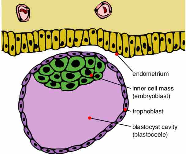
In humans, the blastocyst is formed approximatelyy five days after fertilization. This stage is preceded by the morula. The morula is a solid ball of about 16 undifferentiated, spherical cells. As cell division continues in the morula, the blastomeres change their shape and tightly align themselves against each other. This is called compaction and is likely mediated by cell surface adhesion glycoproteins.
The blastocyst possesses an inner cell mass (ICM), or embryoblast, which subsequently forms the embryo, and an outer layer of cells, or trophoblast, which later forms the placenta. The trophoblast surrounds the inner cell mass and a fluid-filled, blastocyst cavity known as the blastocoele or the blastocystic cavity.
The embryoblast is the source of embryonic stem cells and gives rise to all later structures of the adult organism. The trophoblast combines with the maternal endometrium to form the placenta in eutherian mammals.

Blastocyst
The blastocyst possesses an inner cell mass from which the embryo will develop, and an outer layer of cells, called the trophoblast, which will eventually form the placenta.
Before gastrulation, the cells of the trophoblast become differentiated into two strata. The outer stratum forms a syncytium, which is a layer of protoplasm studded with nuclei that shows no evidence of subdivision into cells (termed the syncytiotrophoblast).
The inner layer, the cytotrophoblast or layer of Langhans, consists of well-defined cells. As already stated, the cells of the trophoblast do not contribute to the formation of the embryo proper; they form the ectoderm of the chorion and play an important part in the development of the placenta.
On the deep surface of the inner cell mass, a layer of flattened cells, called the endoderm, is differentiated and quickly assumes the form of a small sac, called the yolk sac. Spaces appear between the remaining cells of the mass and, by the enlargement and coalescence of these spaces, a cavity called the amniotic cavity is gradually developed.
The floor of this cavity is formed by the embryonic disk, which is composed of a layer of prismatic cells called the embryonic ectoderm. This layer is derived from the inner cell mass and lies in opposition to the endoderm.