 |
This is a file from the Wikimedia Commons. Information from its description page there is shown below.
Commons is a freely licensed media file repository. You can help.
|
 |
This is a featured picture, which means that members of the community have identified it as one of the finest images on the English Wikipedia, adding significantly to its accompanying article. If you have a different image of similar quality, be sure to upload it using the proper free license tag, add it to a relevant article, and nominate it. |
 |
This image was selected as picture of the day on the English Wikipedia for July 4, 2012. |
Summary
| Description |
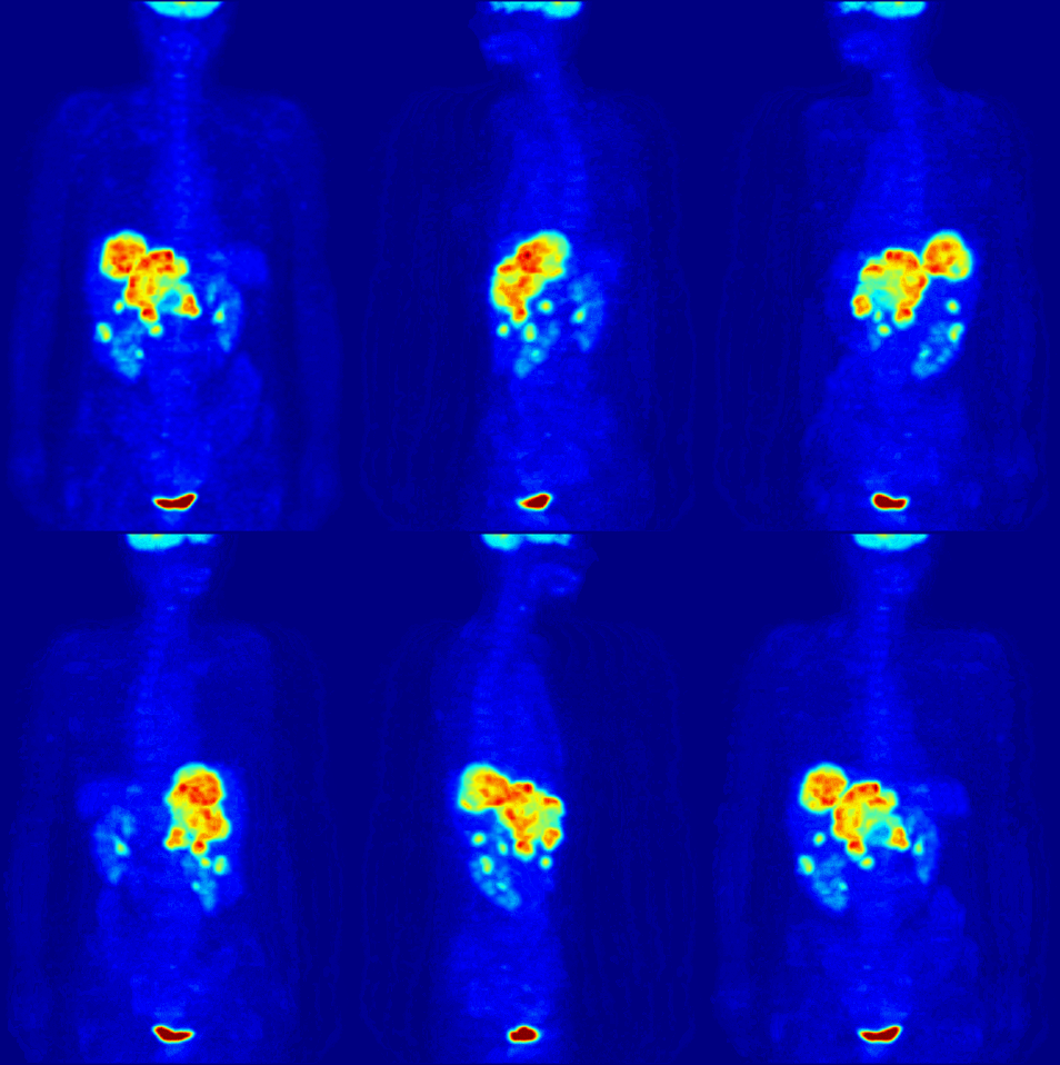
English: Maximum Intensity Projection ( MIP) of a wholebody positron emission tomography ( PET) acquisition of a 79 kg (170 lb) weighting female after intravenous injection of 371 MBq of 18F-FDG (one hour prior measurement). The investigation has been performed as part of a tumor diagnosis prior to applying a radiotherapy (tumor staging step). Besides normal accumulation of the tracer in the heart, bladder, kidneys and brain, liver metastases of a colorectal tumor are clearly visible within the abdominal region of the image.
Deutsch: Maximumintensitätsprojektion ( MIP) einer Ganzkörperaufnahme mittels Positronen-Emissions-Tomographie ( PET). Die Aufnahme zeigt eine 79 kg schwere weibliche Patientin nach intravenöser Injektion von 371 MBq 18F-FDG (eine Stunde vor Messung). Die Untersuchung wurde im Rahmen einer Tumordiagnose vor Anwendung einer Strahlentherapie (sogn. Tumorstaging) durchgeführt. Neben den normalen Anreicherungen des Tracers in Herz, Blase, Nieren und Gehirn, sind auch Lebermetastasen eines kolorektalen Tumor im abdominalen Bereich der Aufnahme auszumachen.
Français : Projection d'intensité maximale ( MIP) d'un corps entier par topographie à émission de positons ( TEP) d'une femme de 79 kg après une injection intraveineuse de 371 MBq de 18F-FDG (une heure avant la mesure). L'étude a été réalisée lors d'un diagnostic de tumeur avant d'appliquer une radiothérapie (étape tumeur). Outre l'accumulation normale du traceur dans le cœur, la vessie, des reins et du cerveau, des métastases hépatiques d'une tumeur colorectale sont clairement visibles dans la région abdominale de l'image.
فارسی: در این تصویر قلب، مثانه، کلیهها، مغز، کبد و نیز متاستاز در سرطان روده بزرگ، کاملا مشخص است.
|
| Date |
22 May 2006 |
| Source |
Own work |
| Author |
Jens Langner ( http://www.jens-langner.de/) |
Permission
( Reusing this file) |
| Public domainPublic domainfalsefalse |
 |
This work has been released into the public domain by its author, I, Jens Langner. This applies worldwide.
In some countries this may not be legally possible; if so:
I, Jens Langner grants anyone the right to use this work for any purpose, without any conditions, unless such conditions are required by law.Public domainPublic domainfalsefalse
|
|
File usage
The following pages on Schools Wikipedia link to this image (list may be incomplete):
SOS Children chose the best bits of Wikipedia to help you learn. SOS Children's Villages helps more than 2 million people across 133 countries around the world. Learn more about child sponsorship.


