 |
This is a file from the Wikimedia Commons. Information from its description page there is shown below.
Commons is a freely licensed media file repository. You can help.
|
Summary
| Description |
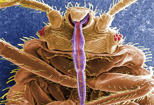
English: ID#: 11739 Description: This digitally-colorized scanning electron micrograph (SEM) revealed some of the ultrastructural morphology displayed on the ventral surface of a bedbug, Cimex lectularius. From this view you can see the insect’s skin piercing mouthparts it uses to obtain its blood meal, as well as a number of its six jointed legs.
|
| Date |
2009 |
| Source |
 |
This media comes from the Centers for Disease Control and Prevention's Public Health Image Library (PHIL), with identification number #11739. Note: Not all PHIL images are public domain; be sure to check copyright status and credit authors and content providers.
|
|
| Author |
Janice Harney Carr, Centre for Disease Control |
Licensing
| Public domainPublic domainfalsefalse |
 |
This image is a work of the Centers for Disease Control and Prevention, part of the United States Department of Health and Human Services, taken or made as part of an employee's official duties. As a work of the U.S. federal government, the image is in the public domain.
česky | Deutsch | English | español | eesti | suomi | français | italiano | македонски | Nederlands | polski | português | slovenščina | 中文 | 中文(简体) | +/−
|
|
File usage
The following pages on Schools Wikipedia link to this image (list may be incomplete):
Schools Wikipedia was created by children's charity SOS Childrens Villages. The world's largest orphan charity, SOS Childrens Villages brings a better life to more than 2 million people in 133 countries around the globe. Have you thought about sponsoring a child?


