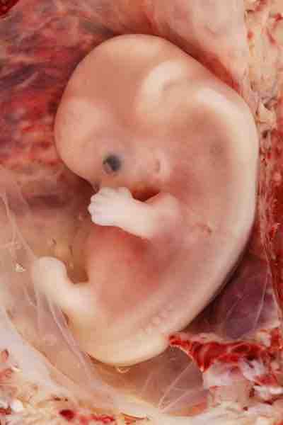
Prenatal development is the process that occurs during the 40 weeks prior to the birth of a child. There are three stages of prenatal development: germinal, embryonic, and fetal. Prenatal development is also organized into three equal trimesters, which do not correspond with the three stages. The first trimester ends with the end of the embryonic stage, the second trimester ends at week 20, and the third trimester ends at birth.
Germinal Stage
The germinal stage is the stage of development that occurs from conception until 2 weeks (implantation). Conception occurs when a sperm fertilizes an egg and forms a zygote. A zygote begins as a one-cell structure that is created when a sperm and egg merge. At the moment of conception, the mother's and father’s DNA are passed on to; the genetic makeup and sex of the future fetus are set at this point. During the first week after conception, the zygote rapidly divides and multiplies, going from a one-cell structure to two cells, then four cells, then eight cells, and so on. This process of cell division is called mitosis. Mitosis is a fragile process, and fewer than one-half of all zygotes survive beyond the first two weeks (Hall, 2004). After 5 days of mitosis there are 100 cells, and after 9 months there are billions of cells. As the cells divide, they become more specialized, forming different organs and body parts. During the germinal stage, the cells necessary for the placenta, umbilical cord, and amniotic fluid will differentiate to form the embryo. The mass of cells has yet to attach itself to the lining of the uterus; once this attachment occurs, the next stage begins.

Embryo
During the germinal stage of prenatal development, the cells necessary for the placenta, umbilical cord, and amniotic fluid will differentiate to form the embryo.
Embryonic Stage
The embryonic stage lasts from implantation (2 weeks) until week 8 of pregnancy. After the zygote divides for about 7–10 days and has 150 cells, it travels down the fallopian tubes and implants itself in the lining of the uterus. Upon implantation, this multi-cellular organism is called an embryo. Now blood vessels grow, forming the placenta. The placenta is a structure connected to the uterus that provides nourishment and oxygen from the woman's body to the developing embryo through the umbilical cord.
During the first week of the embryonic period, the embryonic disk separates into three layers: the ectoderm, mesoderm, and endoderm. The ectoderm is the layer that will become the nervous system and outer skin layers; the mesoderm will become the circulatory system, skeleton, muscles, reproductive system, and inner layer of skin; and the endoderm will become the respiratory system and part of the digestive system, as well as the urinary tract.
The first part of the embryo to develop is the neural tube, which will become the spinal cord and brain. As the nervous system starts to develop, the tiny heart starts to pump blood, and other parts of the body—such as the digestive tract and backbone—begin to emerge. In the second half of this period, growth is very rapid. The eyes, ears, nose, and jaw develop; the heart develops chambers; and the intestines grow.
Fetal Stage
The remainder of prenatal development occurs during the fetal stage, which lasts from week 9 until birth (usually between 38 and 40 weeks). When the organism is about nine weeks old, the embryo is called a fetus. At this stage, the fetus is about the size of a kidney bean and begins to take on the recognizable form of a human being. Between 9 and 12 weeks, reflexes begin to appear and the arm and legs start to move (those first movements won't be felt for a few weeks, however). During this same time, the sex organs begin to differentiate. At about 16 weeks, the fetus is approximately 4.5 inches long. Fingers and toes are fully developed, and fingerprints are visible. By the time the fetus reaches the sixth month of development (24 weeks), it weighs up to 1.4 pounds. Hearing has developed, so the fetus can respond to sounds. The internal organs, including the lungs, heart, stomach, and intestines, have formed enough that a fetus born prematurely at this point has a chance to survive outside of the womb.
Stages of development
During the fetal stage, the brain develops and the body adds size and weight, until the fetus reaches full-term development.
Throughout the fetal stage the brain continues to grow and develop, nearly doubling in size from weeks 16 to 28. Brain growth during this period allows the fetus to develop new behaviors. The cerebral cortex grows larger, and the fetus spends more hours awake. The fetus moves with more coordination, indicating more neural connections within the brain. The nervous system is controlling more bodily functions, and even personality begins to emerge in utero. By 28 weeks, thalamic brain connections form, which mediate sensory input. The fetus can distinguish between voices, and can remember songs and certain sounds after birth. The fetus becomes sensitive to light as well; in fact, if a doctor shines a light on the womb, the baby will attempt to shield his or her eyes. Growth begins to slow around 30 to 32 weeks, but small changes continue until birth.
Around 36 weeks, the fetus is almost ready for birth. It weighs about 6 pounds and is about 18.5 inches long, and by week 37 all of the fetus’s organ systems are developed enough that it could survive outside the uterus without many of the risks associated with premature birth. The fetus continues to gain weight and grow in length until approximately 40 weeks. By then, the fetus has very little room to move around and birth becomes imminent.

Timeline of prenatal development
This chart details prenatal development from conception to birth.