Brain Lateralization
The brain is divided into two halves, called hemispheres. There is evidence that each brain hemisphere has its own distinct functions, a phenomenon referred to as lateralization. The left hemisphere appears to dominate the functions of speech, language processing and comprehension, and logical reasoning, while the right is more dominant in spatial tasks like vision-independent object recognition (such as identifying an object by touch or another nonvisual sense). However, it is easy to exaggerate the differences between the functions of the left and right hemispheres; both hemispheres are involved with most processes. Additionally, neuroplasticity (the ability of a brain to adapt to experience) enables the brain to compensate for damage to one hemisphere by taking on extra functions in the other half, especially in young brains.
Corpus Callosum
The two hemispheres communicate with one another through the corpus callosum. The corpus callosum is a wide, flat bundle of neural fibers beneath the cortex that connects the left and right cerebral hemispheres and facilitates interhemispheric communication. The corpus callosum is sometimes implicated in the cause of seizures; patients with epilepsy sometimes undergo a corpus callostomy, or the removal of the corpus callosum.
The Lobes of the Brain
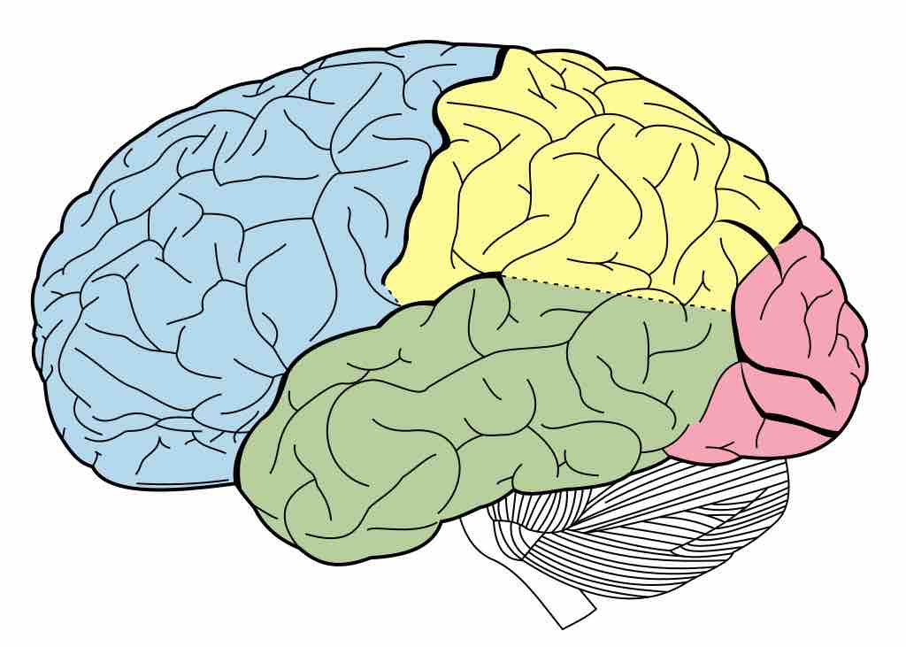
The brain is separated into four lobes: the frontal, temporal, occipital, and parietal lobes.

Lobes of the brain
The brain is divided into four lobes, each of which is associated with different types of mental processes. Clockwise from left: The frontal lobe is in blue at the front, the parietal lobe in yellow at the top, the occipital lobe in red at the back, and the temporal lobe in green on the bottom.
The Frontal Lobe
The frontal lobe is associated with executive functions and motor performance. Executive functions are some of the highest-order cognitive processes that humans have. Examples include:
- planing and engaging in goal-directed behavior;
- recognizing future consequences of current actions;
- choosing between good and bad actions;
- overriding and suppressing socially unacceptable responses;
- determining similarities and differences between objects or situations.
The frontal lobe is considered to be the moral center of the brain because it is responsible for advanced decision-making processes. It also plays an important role in retaining emotional memories derived from the limbic system, and modifying those emotions to fit socially accepted norms.
The Temporal Lobe
The temporal lobe is associated with the retention of short- and long-term memories. It processes sensory input including auditory information, language comprehension, and naming. It also creates emotional responses and controls biological drives such as aggression and sexuality.
The temporal lobe contains the hippocampus, which is the memory center of the brain. The hippocampus plays a key role in the formation of emotion-laden, long-term memories based on emotional input from the amygdala. The left temporal lobe holds the primary auditory cortex, which is important for processing the semantics of speech.
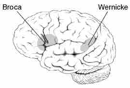
One specific portion of the temporal lobe, Wernicke's area, plays a key role in speech comprehension. Another portion, Broca's area, underlies the ability to produce (rather than understand) speech. Patients with damage to Wernicke's area can speak clearly but the words make no sense, while patients with damage to Broca's area will fail to form words properly and speech will be halting and slurred. These disorders are known as Wernicke's and Broca's aphasia respectively; an aphasia is an inability to speak.

Broca's and Wernicke's areas
The locations of Broca's and Wernicke's areas in the brain. The Broca's area is at the back of the frontal lobe, and the Wernicke's area is roughly where the temporal lobe and parietal lobe meet.
The Occipital Lobe
The occipital lobe contains most of the visual cortex and is the visual processing center of the brain. Cells on the posterior side of the occipital lobe are arranged as a spatial map of the retinal field. The visual cortex receives raw sensory information through sensors in the retina of the eyes, which is then conveyed through the optic tracts to the visual cortex. Other areas of the occipital lobe are specialized for different visual tasks, such as visuospatial processing, color discrimination, and motion perception. Damage to the primary visual cortex (located on the surface of the posterior occipital lobe) can cause blindness, due to the holes in the visual map on the surface of the cortex caused by the lesions.
The Parietal Lobe
The parietal lobe is associated with sensory skills. It integrates different types of sensory information and is particularly useful in spatial processing and navigation. The parietal lobe plays an important role in integrating sensory information from various parts of the body, understanding numbers and their relations, and manipulating objects. Its also processes information related to the sense of touch.
The parietal lobe is comprised of the somatosensory cortex and part of the visual system. The somatosensory cortex consists of a "map" of the body that processes sensory information from specific areas of the body. Several portions of the parietal lobe are important to language and visuospatial processing; the left parietal lobe is involved in symbolic functions in language and mathematics, while the right parietal lobe is specialized to process images and interpretation of maps (i.e., spatial relationships).