Seminiferous tubules, located in the testes, are where meiosis occurs and the gametes (spermatozoa) are created. The seminiferous tubules are formed from primitive sex cords from the gonadal ridge. The epithelium of the tubule consists of tall, columnar cells called Sertoli cells. Between the Sertoli cells are spermatogenic cells, which differentiate through meiosis to become sperm cells. There are two types of seminiferous tubules: convoluted, located toward the lateral side, and straight, as the tubule comes medially to form ducts that will exit the testis.
Efferent Ducts
The efferent ducts connect the rete testis with the initial section of the epididymis. There are two basic types of efferent ductule structure.
1) Multiple entries into the epididymis: This type is seen in most large mammals. In humans and other large mammals, there are approximately 15-20 efferent ducts which occupy nearly one-third of the head of the epididymis.
2) Single entry: This type is seen in most small animals such as rodents. This is characterized by three to six ductules that merge into a single small ductule prior to entering the epididymis.
The ductuli are unilaminar and composed of columnar ciliated and nonciliated (absorptive) cells. The ciliated cells stir the luminal fluids, which may help ensure homogeneous absorption of water from the fluid produced by the testis. This results in an increase in the concentration of luminal sperm. The epithelium is surrounded by a band of smooth muscle that helps to propel the sperm toward the epididymis.

Inside the Human Testes
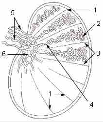
Diagram of cross-section of testes: 1: Testicular septa 2: Convoluted seminiferous tubules 3: Testicular lobules 4: Straight seminiferous tubules 5: Efferent ductules 6: Rete testis.
The ductus (vas) deferens, also called the sperm duct, extend from the epididymis on each side of the scrotum into the abdominal cavity through the inguinal canal, an opening in the abdominal wall for the spermatic cord. The spermatic cord is a connective tissue sheath that contains the vas deferens, testicular blood vessels, and nerves.
The smooth muscle layer of the vas deferens contracts in waves of peristalsis during ejaculation. Two ducts connect the left and right epididymis to the ejaculatory ducts in order to move sperm. In humans, each tube is about 30 centimeters (a foot) long, 3 to 5 mm in diameter, and surrounded by smooth muscle. The sperm are transferred from the vas deferens into the urethra, collecting secretions from the male accessory sex glands such as the seminal vesicles, prostate gland, and bulbourethral glands, which provide the bulk of semen.
The procedure of deferentectomy, also known as a vasectomy, is a method of contraception in which the vas deferens are permanently cut, though in some cases it can be reversed. A modern procedure that does not include cutting the ducts involves injecting an obstructive material into the ductus to block the flow of sperm. Research in male contraception has focused primarily on the vas deferens with the use of the intra-vas device and reversible inhibition of sperm under guidance.

Histiological Representation of Seminferious Tubules
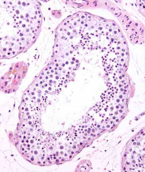
Seminiferous tubule in cross-section (large tubular structure - center of image) with sperm (black, tiny, ovoid bodies furthest from the outer edge of the tubular structure).