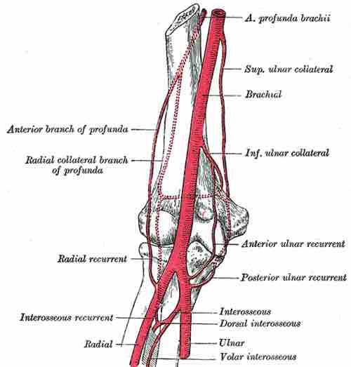
The blood supply of a synovial joint comes from the arteries sharing in anastomosis around the joint. The articular and epiphyseal branches of the neighboring arteries form a periarticular arterial plexus. The articular capsule is highly innervated but avascular (lacking blood and lymph vessels), and receives nutrition from the surrounding blood supply via either the slow process of diffusion or convection, a far more efficient process.
Numerous vessels from this plexus pierce the fibrous capsule and form a rich vascular plexus in the deeper part of the synovial membrane. The blood vessels of the synovial membrane terminate around the articular margins in a fringe of looped anastomoses termed the circulus vasculosus (circulus articularis vasculosus). It supplies the capsule, synovial membrane, and the epiphyses. After epiphyseal fusion in the growth of long bones, communication between the circulosus vasculosus and the end arteries of the metaphysis is established. This minimizes the chances of osteomyelitis in the metaphysis.
The synovial cartilage in the capsule acts somewhat like a sponge. A sponge will absorb fluid, but it will release little of that fluid unless it is squeezed. Exercising the joint, in effect, squeezes the synovial "sponge", allowing gas exchange to occur and nutrients to flow into the cartilage. Flexing and extending the joint alternately squeezes the sponge and releases it to reabsorb more fluid.

Elbow Joint
Diagram of the anastomosis around the elbow joint.