Prenatal Screening Overview
Prenatal diagnosis and prenatal screening are methods for testing for diseases or conditions in a fetus or embryo before it is born. There are three purposes of prenatal diagnosis:
- To enable timely medical or surgical treatment of a condition before (fetal therapy) or after birth.
- To give the parents the chance to decide to abort a fetus with the diagnosed condition.
- To give parents the chance to prepare psychologically, socially, financially, and medically for a baby with a health problem or disability, or for the likelihood of a stillbirth.
Having this information in advance of the birth means that healthcare staff, as well as parents, can better prepare themselves for the delivery of a child with a health problem.
For example, Down Syndrome is associated with cardiac defects that may need intervention immediately upon birth. The other types of conditions commonly screened for include neural tube defects, chromosome abnormalities, genetic diseases, spina bifida, cleft palate, Tay-Sachs disease, sickle cell anemia, thalassemia, cystic fibrosis, and fragile X syndrome.

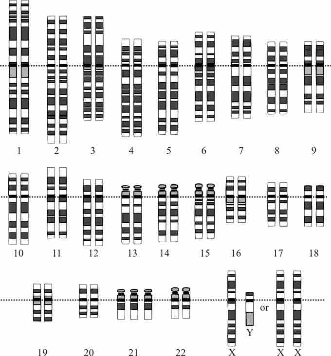
Prenatal diagnosis: Karyotype
This karyotype indicates that the fetus has Down Syndrome as it has three of chromosome 21 instead of two.
Fetal screening is also done to determine characteristics that are not generally considered birth defects, such as determining the sex of the offspring. The primary method for sex determination is prenatal ultrasound. Cell-free fetal DNA testing is also possible, wherein the maternal blood is analyzed to detect the small amount of free circulating fetal DNA.
Invasive and Noninvasive Prenatal Screening
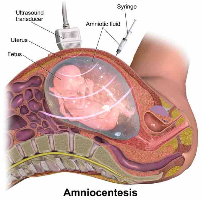
Diagnostic prenatal testing can be done by invasive or noninvasive methods. An invasive method involves probes or needles being inserted into the uterus, e.g., amniocentesis (that can be done from about 14 weeks gestation up to about 20 weeks), and chorionic villus sampling (that can be done earlier: between 9.5 and 12.5 weeks gestation).
Chorionic villus sampling is associated with slightly more risk to the fetus. Since chorionic villus sampling is performed earlier in the pregnancy than amniocentesis, typically during the first trimester, it can reasonably be expected that there will be a higher rate of miscarriage after chorionic villus sampling than after amniocentesis.
Non-invasive techniques include examinations of the woman's womb through ultrasonography and maternal serum screens. Blood tests for select trisomies based on detecting fetal DNA present in maternal blood are available. Technologies to perform prenatal testing in a noninvasive manner reduce the risk of pregnancy loss that can occur with other methods.
These include the use of maternal blood and DNA sequencing methods such as next-generation sequencing to determine the presence of genetic defects. If an elevated risk of chromosomal or genetic abnormality is indicated by a noninvasive screening test, a more invasive technique may be employed to gather more information. In the case of neural tube defects, a detailed ultrasound can non-invasively provide a definitive diagnosis.
Because of the risk of miscarriage and fetal damage associated with amniocentesis and chorionic villus sampling procedures, many women prefer to first undergo screening so they can find out if the fetus' risk of birth defects is high enough to justify the risks of invasive testing. Screening tests yield a risk score that represents the chance that a baby has the birth defect.
The most common threshold for high risk is 1:270. A risk score of 1:300 would be considered low-risk by many physicians. However, the trade-off between the risk of a birth defect and the risk of complications from invasive testing is relative and subjective. Some parents may decide that even a 1:1,000 risk of birth defects warrants an invasive test, while others would not opt for an invasive test even if they had a 1:10 risk score.
The American Congress of Obstetricians and Gynecologists guidelines currently recommend that all pregnant women, regardless of age, be offered invasive testing to obtain a definitive diagnosis of certain birth defects. Therefore, most physicians offer diagnostic testing to all their patients, with or without prior screening, and let the patient decide. Invasive testing is warranted in the following cases:
- Women over the age of 35.
- Women who have previously had premature babies or babies with a birth defect.
- Women who have high blood pressure, lupus, diabetes, asthma, or epilepsy.
- Women who have family histories or ethnic backgrounds prone to genetic disorders, or whose partners have these.
- Women who are pregnant with twins or more.
- Women who have miscarried.

Amniocentesis
An illustration of the amniocentesis procedure.
Determination of Gestational Age
At about 6 weeks of pregnancy, an ultrasound dating scan may be offered to help confirm the gestational age of the embryo and check if a single or twin pregnancy exists, but such a scan is unable to detect common abnormalities. Around weeks 10–11, a nuchal thickness scan (NT) may be offered, which can be combined with blood tests that correlate with chromosomal abnormalities.
The results of the blood tests are then combined with the NT ultrasound measurements, maternal age, and gestational age of the fetus to yield a risk score for Down syndrome, trisomy 18, and trisomy 13. Other testing may combine a first trimester blood test with one done in the second trimester to determine if further action needs to be taken.