The basal ganglia (or basal nuclei, ) are a group of nuclei of varied origin in the brains of vertebrates that act as a cohesive functional unit. They are situated at the base of the forebrain and are strongly connected with the cerebral cortex, thalamus, and other brain areas. The basal ganglia are associated with a variety of functions, including voluntary motor control, procedural learning relating to routine behaviors or "habits" such as bruxism, eye movements, and cognitive, emotional functions. Currently, popular theories implicate the basal ganglia primarily in action selection, that is, the decision of which several possible behaviors to execute at a given time. Experimental studies show that the basal ganglia exert an inhibitory influence on a number of motor systems, and that a release of this inhibition permits a motor system to become active. The "behavior switching" that takes place within the basal ganglia is influenced by signals from many parts of the brain, including the prefrontal cortex, which plays a key role in executive functions.

The Basal Ganglia
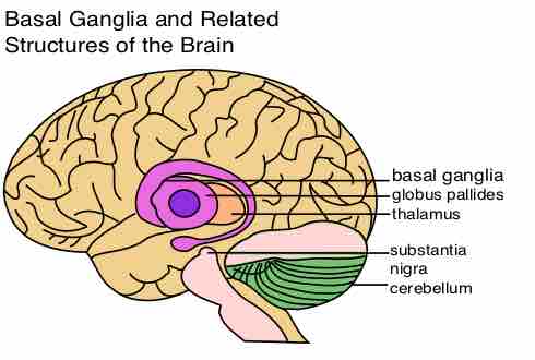
The basal nuclei are often referred to as the basal ganglia. The main components of the basal nuclei are labeled in purple.
The main components of the basal ganglia are:
- the striatum, or neostriatum (composed of the caudate and putamen). The largest component, the striatum, receives input from many brain areas, but sends output only to other components of the basal ganglia.
- the globus pallidus, or pallidum (composed of globus pallidus externa (GPe) and globus pallidus interna (GPi)). The pallidum receives its most important input from the striatum (either directly or indirectly), and sends inhibitory output to a number of motor-related areas, including the part of the thalamus that projects to the motor-related areas of the cortex.
- the substantia nigra (composed of both substantia nigra pars compacta (SNc) and substantia nigra pars reticulata (SNr)). One part of substantia nigra, the reticulata (SNr), functions similarly to the pallidum, and another part (compacta or SNc) provides the source of the neurotransmitter dopamine's input to the striatum.
- the subthalamic nucleus (STN). The subthalamic nucleus (STN) receives input mainly from the striatum and cortex, and projects to a portion of the pallidum (interna portion or GPi). Each of these areas has a complex internal anatomical and neurochemical organization.
The basal ganglia play a central role in a number of neurological conditions, including several movement disorders. The most notable are Parkinson's disease, which involves degeneration of the melanin-pigmented dopamine-producing cells in the substantia nigra pars compacta (SNc), and Huntington's disease, which primarily involves damage to the striatum. Basal ganglia dysfunction is also implicated in some other disorders of behavior control such as the Tourette's syndrome, ballismus (particularly hemibalismus), obsessive–compulsive disorder (OCD), and Wilson's disease (Hepatolenticular degeneration). With the exception of Wilson's disease and hemiballismus, the neuropathological mechanisms underlying diseases of ganglia such as Parkinsons' and Huntington's are not very well understood or are at best still developing theories.

The Basal Ganglia
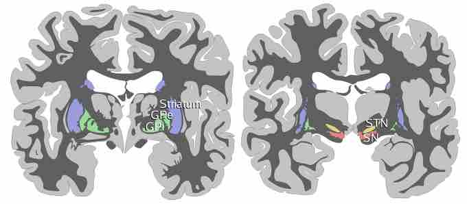
Two schematic drawings of coronal sections of human brain labelling the basal ganglia. Blue=striatum, green=globus pallidus (external and internal segments), yellow=subthalamic nucleus, red=substantia nigra (pars reticulata and pars compacta). The right section is the deeper one, closer to the back of the head.