Autonomic ganglia are clusters of neuronal cell bodies and their dendrites. They are essentially a junction between autonomic nerves originating from the central nervous system and autonomic nerves innervating their target organs in the periphery.
The dorsal root ganglia lie along the vertebral column by the spine and develop in the embryo from neural crest cells, not neural tube. Therefore, the spinal ganglia can be regarded as gray matter of the spinal cord that became translocated to the periphery.
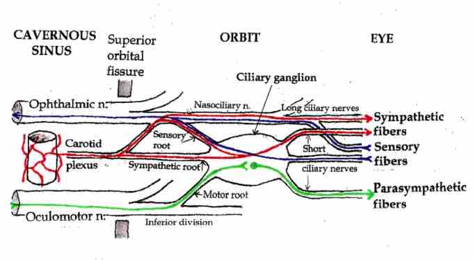
The two main categories are: sympathetic ganglia and parasympathetic ganglia. An example of parasympathetic ganglion is the ciliary ganglion, involved in pupil constriction and accommodation. A depiction of all the parasympathetic ganglia in the head and neck is shown in the following illustration.

Ciliary ganglion
The pathways of the ciliary ganglion include sympathetic neurons (red), parasympathetic neurons (green), and sensory neurons (blue).
Parasympathetic ganglia of the head
Parasympathetic ganglia of the head (shown as red circles) help supply all parasympathetic innervation to the head and neck.

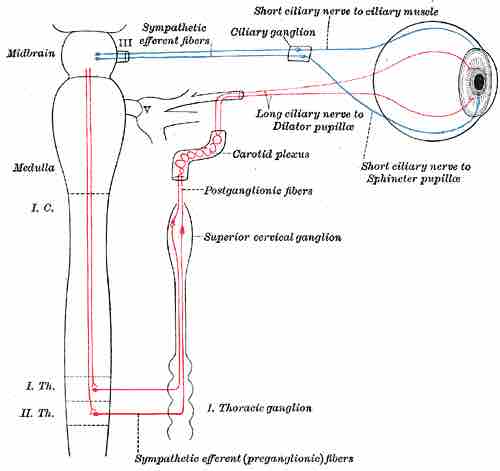
Anatomy of an autonomic ganglion
The sympathetic connections of the ciliary and superior cervical ganglia are shown in this digram. The postganglionic fibers travel from the ganglion to the effector organ.
Dorsal Root Ganglia
A dorsal root ganglion (or spinal ganglion) is a nodule on a dorsal root of the spine that contains the cell bodies of nerve cells (neurons) that carry signals from the sensory organs towards the appropriate integration center.
Nerves that carry signals towards the brain are known as afferent nerves. The axons of dorsal root ganglion neurons are known as afferents. In the peripheral nervous system, afferents refer to the axons that relay sensory information into the central nervous system (i.e., the brain and the spinal cord).
These neurons are of the pseudo-unipolar type, meaning they have an axon with two branches that act as a single axon, often referred to as a distal process and a proximal process.
Unlike the majority of neurons found in the central nervous system, an action potential in a dorsal root ganglion neuron may initiate in the distal process in the periphery, bypass the cell body, and continue to propagate along the proximal process until reaching the synaptic terminal in the dorsal horn of the spinal cord.
The distal section of the axon may either be a bare nerve ending or encapsulated by a structure that helps relay specific information to a nerve. The nerve endings of dorsal root ganglion neurons have a variety of sensory receptors that are activated by mechanical, thermal, chemical, and noxious stimuli.
In these sensory neurons, a group of ion channels thought to be responsible for somatosensory transduction have been identified. For example, a Meissner's corpuscle or Pacinian corpuscle may encapsulate the nerve ending, rendering the distal process sensitive to mechanical stimulation, such as stroking or vibration, respectively.
Sympathetic Ganglia
Sympathetic ganglia are the ganglia of the sympathetic nervous system. They deliver information to the body about stress and impending danger, and are responsible for the familiar fight-or-flight response. They contain approximately 20,000–30,000 nerve cell bodies and are located close to and on either side of the spinal cord in long chains.
Sympathetic ganglia are the tissue from which neuroblastoma tumors arise. The bilaterally symmetric sympathetic chain ganglia—also called the paravertebral ganglia—are located just ventral and lateral to the spinal cord. The chain extends from the upper neck down to the coccyx, forming the unpaired coccygeal ganglion.
Preganglionic nerves from the spinal cord create a synapse end at one of the chain ganglia, and the postganglionic fiber extends to an effector, typically a visceral organ in the thoracic cavity. There are usually 21 or 23 pairs of these ganglia: three in the cervical region, 12 in the thoracic region, four in the lumbar region, four in the sacral region and a single, unpaired ganglion lying in front of the coccyx called the ganglion impar.
Neurons of the collateral ganglia, also called the prevertebral ganglia, receive input from the splanchnic nerves and innervate organs of the abdominal and pelvic region. These include the celiac ganglia, superior mesenteric ganglia, and inferior mesenteric ganglia.
Parasympathetic Ganglia
Parasympathetic ganglia are the autonomic ganglia of the parasympathetic nervous system. Most are small terminal ganglia or intramural ganglia, so named because they lie near or within (respectively) the organs they innervate. The exceptions are the four paired parasympathetic ganglia of the head and neck.
Efferent parasympathetic nerve signals are carried from the central nervous system to their targets by a system of two neurons. The first neuron in this pathway is referred to as the preganglionic or presynaptic neuron. Its cell body sits in the central nervous system and its axon usually extends to a ganglion somewhere else in the body, where it synapses with the dendrites of the second neuron in the chain.
This second neuron is referred to as the postganglionic or postsynaptic neuron. The axons of presynaptic parasympathetic neurons are usually long. They extend from the CNS into a ganglion that is either very close to or embedded in their target organ. As a result, the postsynaptic parasympathetic nerve fibers are very short.