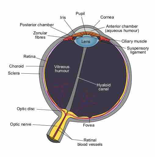
The human eye is an organ that reacts to light in many circumstances. As a conscious sense organ the human eye allows vision; rod and cone cells in the retina allow conscious light perception and vision, including color differentiation and the perception of depth. The human eye can distinguish about 10 million colors. A model of the human eye can be seen in .

Schematic Diagram of the Human Eye
Structure of the eye and closeup of the retina.
The retina of human eye has a static contrast ratio of around 100:1 (about 6.5 f-stops). As soon as the eye moves, it re-adjusts its exposure, both chemically and geometrically, by adjusting the iris (which regulates the size of the pupil). Initial dark adaptation takes place in approximately four seconds of profound, uninterrupted darkness; full adaptation, through adjustments in retinal chemistry, is mostly complete in thirty minutes. Hence, a dynamic contrast ratio of about 1,000,000:1 (about 20 f-stops) is possible. The process is nonlinear and multifaceted, so an interruption by light starts the adaptation process over again. Full adaptation is dependent on good blood flow (thus dark adaptation may be hampered by poor circulation, and vasoconstrictors like tobacco).
The eye includes a lens not dissimilar to lenses found in optical instruments (such as cameras). The same principles can be applied. The pupil of the human eye is its aperture. The iris is the diaphragm that serves as the aperture stop. Refraction in the cornea causes the effective aperture (the entrance pupil) to differ slightly from the physical pupil diameter. The entrance pupil is typically about 4 mm in diameter, although it can range from 2 mm (f/8.3) in a brightly lit place to 8 mm (f/2.1) in the dark. The latter value decreases slowly with age; older people's eyes sometimes dilate to not more than 5-6mm.
The approximate field of view of an individual human eye is 95° away from the nose, 75° downward, 60° toward the nose, and 60° upward, allowing humans to have an almost 180-degree forward-facing horizontal field of view. With eyeball rotation of about 90° (head rotation excluded, peripheral vision included), horizontal field of view is as high as 170°. About 12–15° temporal and 1.5° below the horizontal is the optic nerve or blind spot which is roughly 7.5° high and 5.5° wide.