Magnification is the process of enlarging something only in appearance, not in physical size. This enlargement is quantified by a calculated number also called "magnification. " The term magnification is often confused with the term "resolution," which describes the ability of an imaging system to show detail in the object that is being imaged. While high magnification without high resolution may make very small microbes visible, it will not allow the observer to distinguish between microbes or sub-cellular parts of a microbe. In reality, therefore, microbiologists depend more on resolution, as they want to be able to determine differences between microbes or parts of microbes. However, to be able to distinguish between two objects under a microscope, a viewer must first magnify to a point at which resolution becomes relevant .

Aging Tissue and Vision Loss
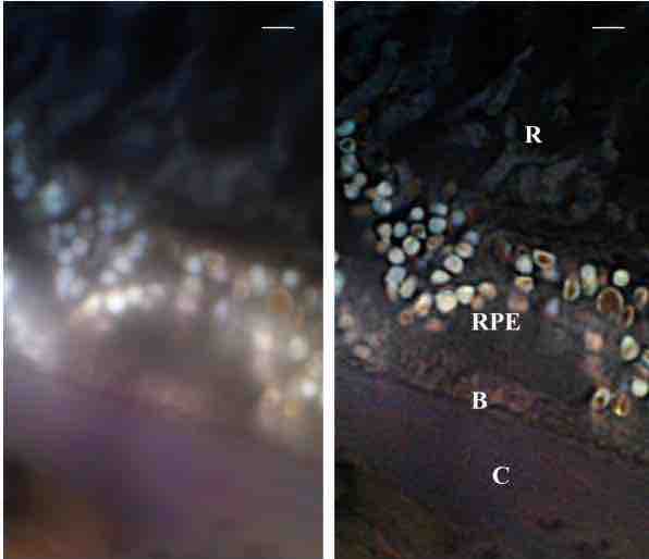
These are micrographs of a section of a human eye. Using computer algorithms and other technology the panel on the right has a higher resolution and is therefore clearer. It should be noted that both panels are at the same magnification, yet the panel on the right has a higher resolution and gives more information on the sample. The labels represent various parts of the human eye: Bruch membrane (B); choroid (C); retinal pigment epithelium (RPE); and retinal rod cells (R). The scale bar is 2um.
Resolution depends on the distance between two distinguishable radiating points. A microscopic imaging system may have many individual components, including a lens and recording and display components. Each of these contributes to the optical resolution of the system, as will the environment in which the imaging is performed. Real optical systems are complex, and practical difficulties often increase the distance between distinguishable point sources.
At very high magnifications with transmitted light, point objects are seen as fuzzy discs surrounded by diffraction rings. These are called Airy disks. The resolving power of a microscope is taken as the ability to distinguish between two closely spaced Airy disks (or, in other words, the ability of the microscope to distinctly reveal adjacent structural detail). It is this effect of diffraction that limits a microscope's ability to resolve fine details. The extent and magnitude of the diffraction patterns are affected by the wavelength of light (λ), the refractive materials used to manufacture the objective lens, and the numerical aperture (NA) of the objective lens. There is therefore a finite limit beyond which it is impossible to resolve separate points in the objective field. This is known as the diffraction limit.