Concept
Version 8
Created by Boundless
Magnification and Resolution

Aging Tissue and Vision Loss
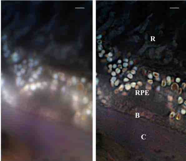
These are micrographs of a section of a human eye. Using computer algorithms and other technology the panel on the right has a higher resolution and is therefore clearer. It should be noted that both panels are at the same magnification, yet the panel on the right has a higher resolution and gives more information on the sample. The labels represent various parts of the human eye: Bruch membrane (B); choroid (C); retinal pigment epithelium (RPE); and retinal rod cells (R). The scale bar is 2um.
Source
Boundless vets and curates high-quality, openly licensed content from around the Internet. This particular resource used the following sources:
"Opthalmology AMD Super Resolution Cremer."
http://commons.wikimedia.org/wiki/File:Opthalmology_AMD_Super_Resolution_Cremer.png
Wikimedia
CC BY-SA 3.0.