Atomic Methods in Bacterial Analysis
Crystallography is the scientific study of the arrangement of atoms in a solid. The field has greatly advanced with the development of x-ray diffraction methods, where the matter analyzed is usually in its crystal form. Nuclear magnetic resonance spectroscopy and x-ray crystallography have become the methods of choice for understanding three-dimensional protein structures.

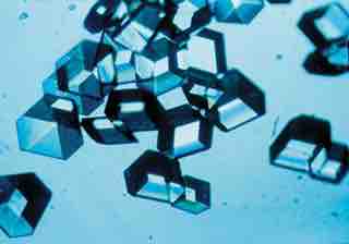
Insulin crystals
insulin crystals
METHODS OF CRYSTALLOGRAPHY
- X-ray crystallography is the primary method for determining the molecular conformation of biological macromolecules, particularly proteins and nucleic acids such as DNA and RNA. Indeed, the double-helical structure of DNA was deduced from crystallographic data.
- Neutron crystallography is often used to help refine structures obtained by x-ray methods or to solve a specific bond; the methods are often viewed as complementary, as x-rays are sensitive to electron positions and scatter most strongly off heavy atoms, while neutrons are sensitive to nucleus positions and scatter strongly off many light isotopes, including hydrogen and deuterium.
- Electron crystallography has been used to determine some protein structures, most notably membrane proteins and viral capsids.
ANALYSIS OF DATA
Studies of protein crystallography help determine the three dimensional structure of proteins and analyze their function alone or within multimolecular assemblies. The structure-function analysis is completed by biochemical and biophysical studies in solution. The protocol for completing a successful crystallographic analysis requires production of proteins (cloning, mutagenesis, bacterial culture, etc.), purification of recombinant proteins (such as chromatography of affinity and gel filtration), enzymatic tests and inhibition measurement (spectrophotometry), crystallization, x-rays crystallography and structural analysis, interactions determination (microcalorimetry, fluorescence, BIAcore), conformational analyses (circular dichroism, ultracentrifugation, light scattering), modifications analysis (mass spectrometry), bioinformatics, and molecular modelisation.
The Protein Data Bank (PDB) is a freely accessible repository documenting the structures of proteins and other biological macromolecules. It stores information about crystals and crystal structures. Computer programs like RasMol or Pymol can be used to visualize biological molecular structures.