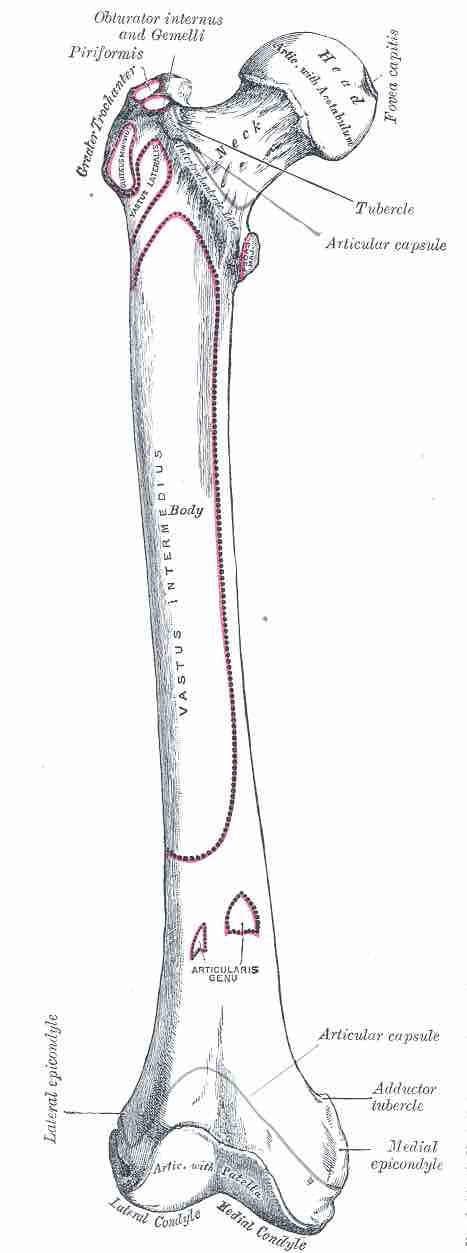
The femur or thigh bone is found in the upper leg and is the longest bone in the body. The femur articulates proximally with the acetabulum of the pelvis to form the hip joint, and distally with the tibia and patella to form the knee joint.

Femur
The anterior surface of the femur with parts labeled
Proximal
Proximally, the femur exhibits four key regions. The femoral head projects medially and superiorly and articulates with the acetabulum of the pelvis to form the hip joint. Immediately lateral to the head is the neck that connects the head with the shaft. It is narrower than the head to permit a greater range of movement at the hip joint.
Located superiorly on the main shaft, lateral to the joining of the neck, the greater trochanter is a projection to which the abductor and lateral rotator muscles of the leg attach.
Also located on the main shaft, but inferiorly to the neck joint, is the lesser trochanter. A much smaller projection than the greater trochanter, the psoas major and iliacus muscles attach here.
Shaft
The shaft descends in a slightly medial direction that is designed to bring the knees closer to the body’s center of gravity, increasing stability. Due to the widening of the female pelvis this angle is greater in women and can lead to increased knee instability.
Two key features of the shaft are the proximal gluteal tuberosity to which the gluteus maximus attaches, and the distal adductor tubercle to which the adductor magnus attaches.
Distal
Distally, the femur exhibits five key regions. Two rounded regions, termed the medial and lateral condyles, articulate with the tibia at the most anterior projection of the patella.
Between the two condyles lies the intercondylar fossa, a depression in which key knee ligaments attach; this significantly strengthens the knee joint and protects it against torsional damage.
Finally, the two epicondyles, the medial and lateral, lie immediately proximal to the condyles; they are also regions where key internal knee ligaments attach.