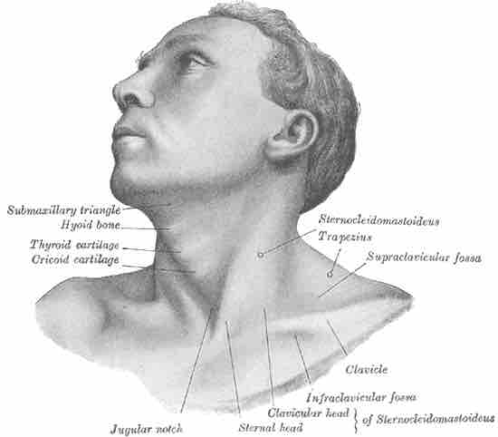
The hyoid bone is a horseshoe shaped bone found in the neck. Located anteriorly between the mandible and the thyroid cartilage, the hyoid bone protects the esophagus and also facilitates the wide range of muscle activity required for speaking and swallowing. It is visible upon extension of the neck.

Position of the hyoid bone in the neck
This image shows where the hyoid bone is located.
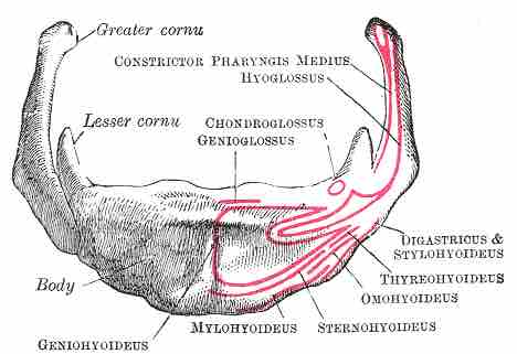
The hyoid bone consists of a central body and two pairs of cornua, or horns, termed greater and lesser cornua. The greater horns project backwards from the body and provide a platform for key muscles and ligaments to attach to including the stylohyoid and throhyoid muscles.
The lesser horns are two smaller eminences which project superiorly to the join between the greater cornua and body and are attached to the body by fibrous tissue. As with the greater cornua the lesser cornua provide a platform for muscle and ligament attachment specifically for the stylohyoid ligament.

Hyoid Bone
Anterior view of hyoid bone with sections labeled.
The hyoid ossifies towards the end of fetal development, commencing in the greater cornua before completing in the body shortly after birth. The greater cornua and body are initially connected by fibrous material although this ossifies towards middle age.