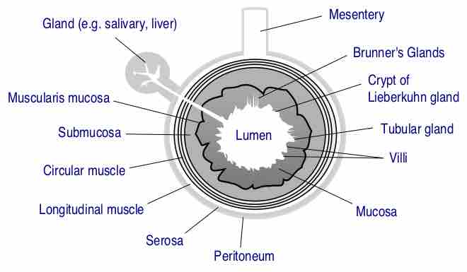
The GI tract is composed of four layers. Each layer has different tissues and functions. From the inside out they are called: mucosa, submucosa, muscularis, and serosa . The submucosa is relatively thick, highly vascular, and serves the mucosa. The absorbed elements that pass through the mucosa are picked up from the blood vessels of the submucosa. The submucosa also has glands and nerve plexuses. The submucosa lies under the mucosa and consists of fibrous connective tissue, separating the mucosa from the next layer, the muscularis externa. The muscularis in the stomach differs from that of other GI organs in that it has three layers of muscle instead of two. Under these muscle layers is the adventitia, layers of connective tissue continuous with the omenta.
Layers of stomach lining.
Stomach. (Serosa is labeled at far right, and is colored yellow. )

General Structure of the Gut Wall
General structure of the gut wall.
The submucosa consists of a dense irregular layer of connective tissue with large blood vessels, lymphatics, and nerves branching into the mucosa and muscularis externa. It contains Meissner's plexus, an enteric nervous plexus, situated on the inner surface of the muscularis externa. In the gastrointestinal tract, the submucosa is the layer of dense irregular connective tissue or loose connective tissue that supports the mucosa. It also joins the mucosa to the bulk of underlying smooth muscle (fibers running circularly within layer of longitudinal muscle). Blood vessels, lymphatic vessels, and nerves (all supplying the mucosa) will run through here. Tiny parasympathetic ganglia are scattered around forming the submucosal plexus (or "Meissner's plexus") where preganglionic parasympathetic neurons synapse with postganglionic nerve fibers that supply the muscularis mucosae.