Concept
Version 13
Created by Boundless
Anatomy of the Heart

The Mammalian Heart
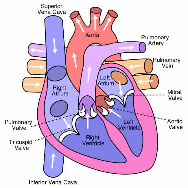
The position of valves ensures proper directional flow of blood through the cardiac interior. Note the difference in the thickness of the muscled walls of the atrium and the left and right ventricle.
This diagram of the heart indicates the superior vena cava, aorta, pulmonary artery, pulmonary vein, left and right atria, mitral valve, aortic valve, left and right ventricles, inferior vena cava, tricuspid valve, and pulmonary valve.
Source
Boundless vets and curates high-quality, openly licensed content from around the Internet. This particular resource used the following sources:
"Diagram of the human heart (cropped)."
http://en.wikipedia.org/wiki/File:Diagram_of_the_human_heart_(cropped).svg%23globalusage
Wikipedia
CC BY-SA 3.0.