Concept
Version 14
Created by Boundless
Preganglionic Neurons

Parasympathetic ganglia of the head
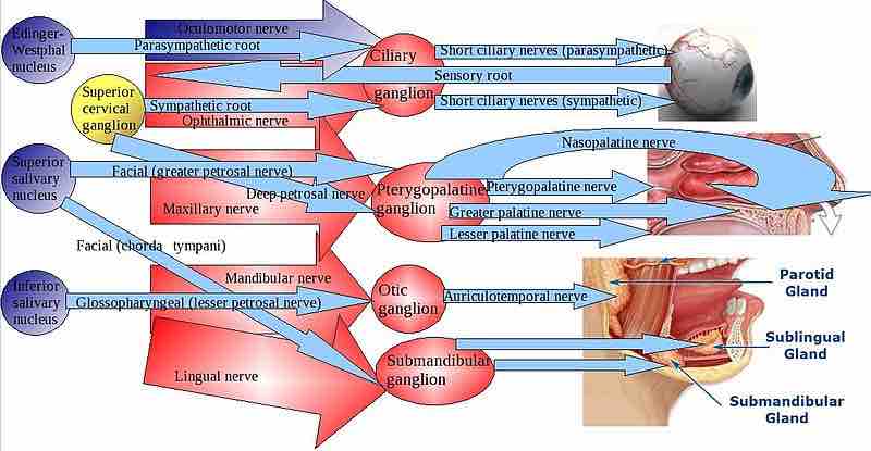
The parasympathetic division has craniosacral outflow, meaning that the neurons begin at the cranial nerves (CN3, CN7, CN9, CN10) and the sacral (S2–S4) spinal cord. Pre- and post-ganglionic fibers and targets are depicted.
This is a diagram of the parasympathetic division of the head. It depicts craniosacral outflow, meaning that the neurons begin at the cranial nerves (CN3, CN7, CN9, CN10) and the sacral (S2-S4) spinal cord. The pre- and post-ganglionic fibers and targets are depicted for the eyes, nose, and mouth.
Source
Boundless vets and curates high-quality, openly licensed content from around the Internet. This particular resource used the following sources:
"Parasympathetic head ganglia."
http://en.wikipedia.org/wiki/File:Parasympathetic_head_ganglia.jpg
Wikipedia
CC BY-SA 3.0.