Brain
The brain is the part of the central nervous system that is contained in the cranial cavity of the skull. It includes the cerebral cortex, limbic system, basal ganglia, thalamus, hypothalamus, and cerebellum.
Cerebral Cortex
The outermost part of the brain is a thick piece of nervous system tissue called the cerebral cortex, which is folded into hills called gyri (singular: gyrus) and valleys called sulci (singular: sulcus). The cortex is composed of two hemispheres, right and left, which are separated by a large sulcus. A thick fiber bundle, the corpus callosum, connects the two hemispheres, allowing information to be passed from one side to the other. Although there are some brain functions that are localized more to one hemisphere than the other, the functions of the two hemispheres are largely redundant. In fact, sometimes (very rarely) an entire hemisphere is removed to treat severe epilepsy. While patients do suffer some deficits following the surgery, they can have surprisingly few problems, especially when the surgery is performed on children who have relatively-undeveloped nervous systems.
In other surgeries to treat severe epilepsy, the corpus callosum is cut instead of removing an entire hemisphere. This causes a condition called split-brain syndrome, which gives insights into unique functions of the two hemispheres. For example, when an object is presented to patients' left visual fields, they may be unable to verbally name the object (and may claim not to have seen an object at all). This is because the visual input from the left visual field crosses and enters the right hemisphere and is unable to signal to the speech center, which generally is found in the left side of the brain. Remarkably, if a split-brain patient is asked to pick up a specific object out of a group of objects with the left hand, the patient will be able to do so, but will still be unable to vocally identify it.
The Four Brain Lobes
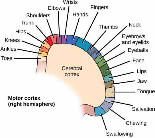
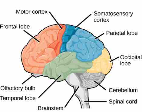
Each hemisphere of the mammalian cerebral cortex can be broken down into four functionally- and spatially-defined lobes: frontal, parietal, temporal, and occipital . The frontal lobe is located at the front of the brain, over the eyes. This lobe contains the olfactory bulb, which processes smells. The frontal lobe also contains the motor cortex, which is important for planning and implementing movement. Areas within the motor cortex map to different muscle groups; there is some organization to this map . For example, the neurons that control movement of the fingers are next to the neurons that control movement of the hand. Neurons in the frontal lobe also control cognitive functions such as maintaining attention, speech, and decision-making. Studies of humans who have damaged their frontal lobes show that parts of this area are involved in personality, socialization, and assessing risk.

Motor cortex control of muscle movement
Different parts of the motor cortex control different muscle groups. Muscle groups that are neighbors in the body are generally controlled by neighboring regions of the motor cortex as well. For example, the neurons that control shoulder movement are near the neurons that control elbow movement, which are themselves next to those that control wrist movement.

Lobes of the cerebral cortex
The human cerebral cortex includes the frontal, parietal, temporal, and occipital lobes, each of which is involved in a different higher function.
The parietal lobe is located at the top of the brain. Neurons in the parietal lobe are involved in speech and reading. Two of the parietal lobe's main functions are processing somatosensation (touch sensations such as pressure, pain, heat, cold) and processing proprioception (the sense of how parts of the body are oriented in space). The parietal lobe contains a somatosensory map of the body similar to the motor cortex.
The occipital lobe is located at the back of the brain. It is primarily involved in vision: seeing, recognizing, and identifying the visual world.
The temporal lobe is located at the base of the brain by the ears. It is primarily involved in processing and interpreting sounds. It also contains the hippocampus (Greek for "seahorse", which is what it resembles), a structure that processes memory formation. The role of the hippocampus in memory was partially determined by studying one famous epileptic patient, HM, who had both sides of his hippocampus removed in an attempt to cure his epilepsy. His seizures went away, but he could no longer form new memories (although he could remember some facts from before his surgery and could learn new motor tasks).